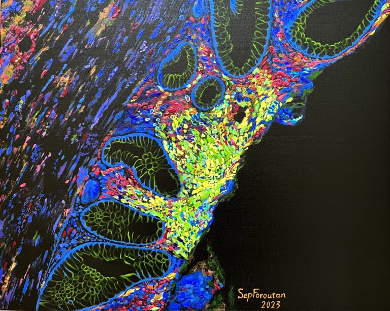
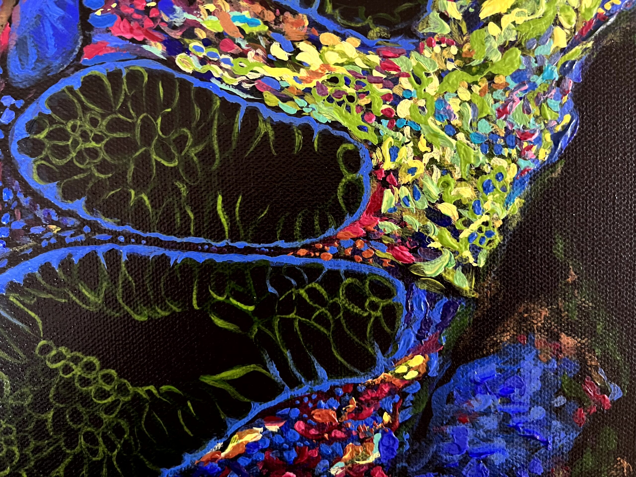
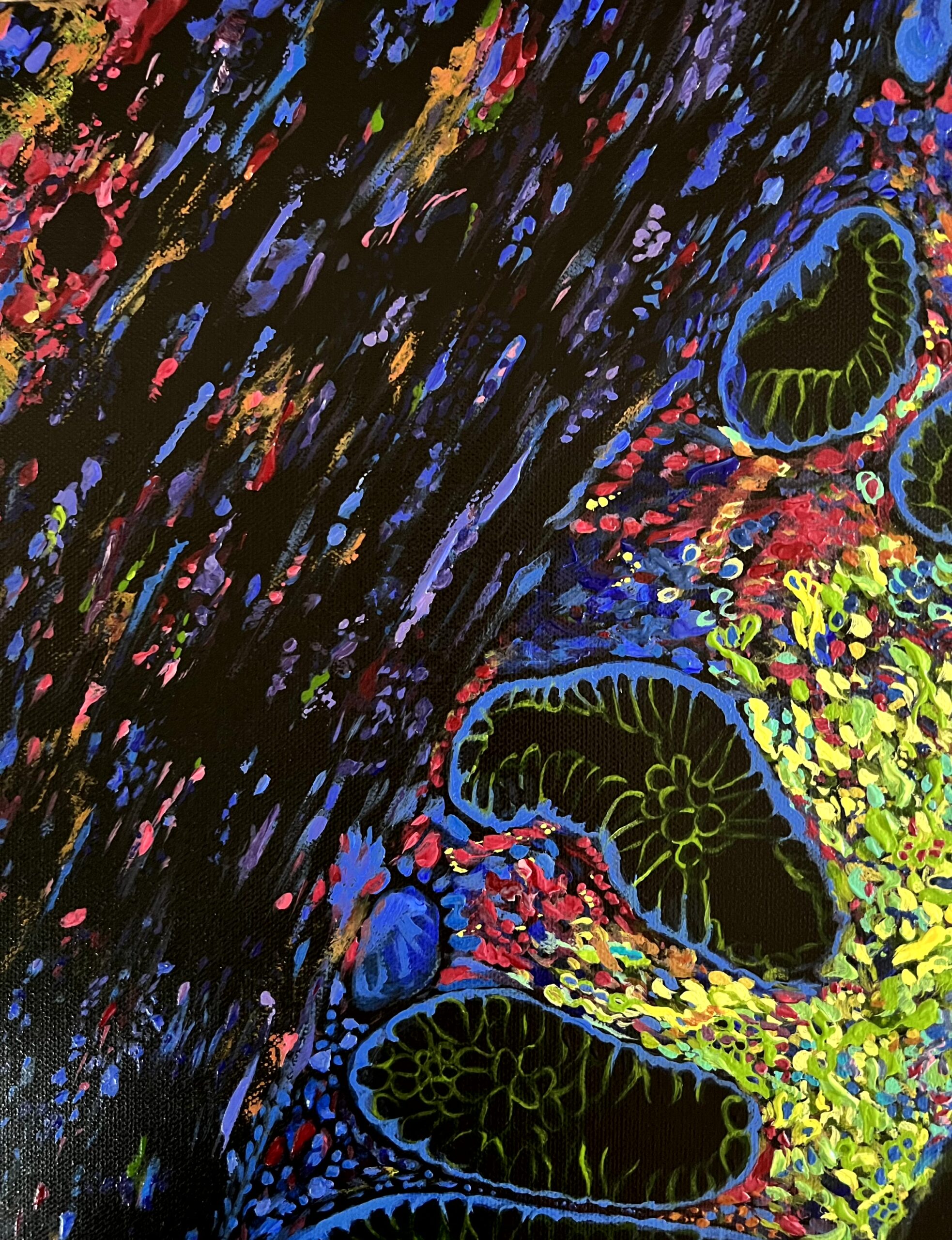
Swirl with Rival!
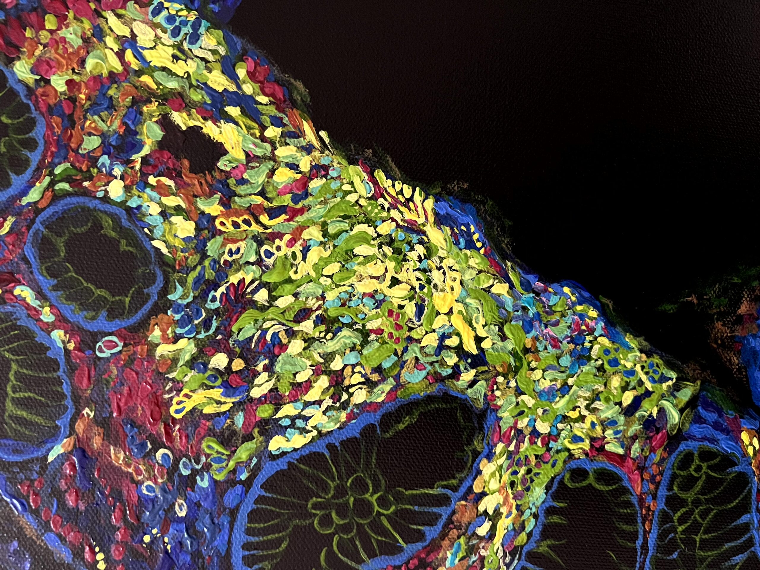
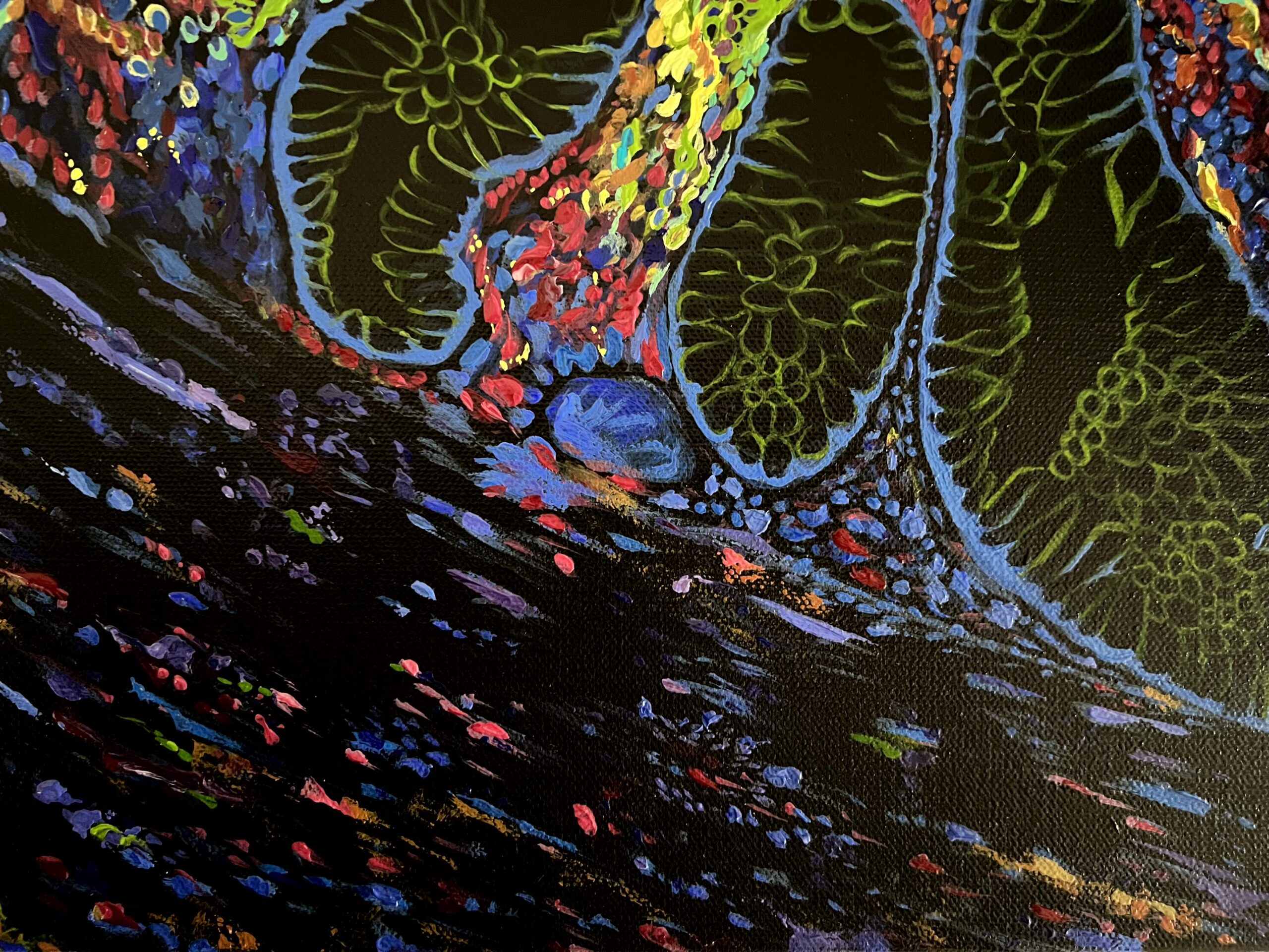
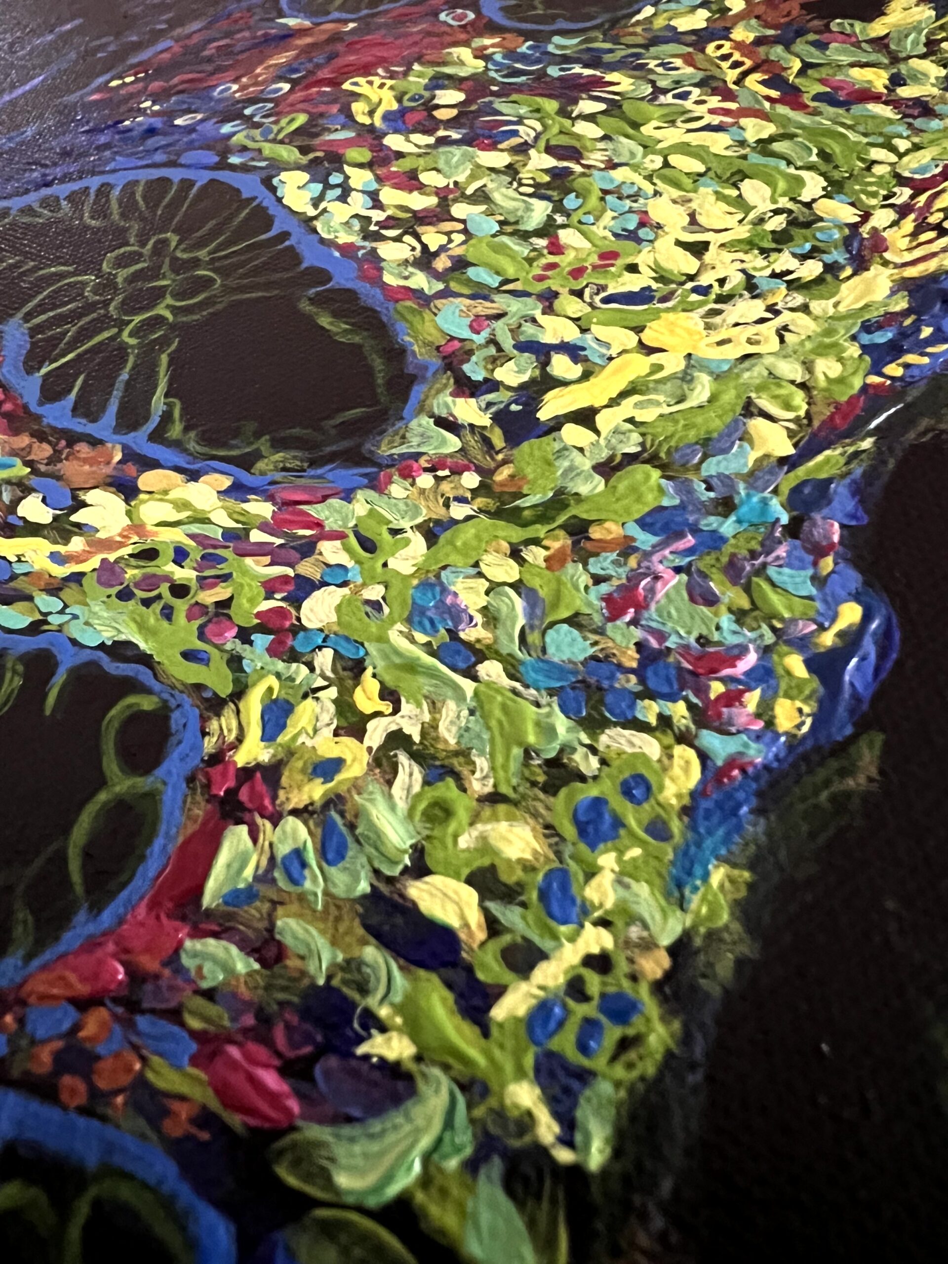
This painting illustrates aggregates of immune cells in rectal cancer! In cancer samples, we often see the up-regulation of a protein called PD-L1 (shown in yellow) which acts as a brake to inhibits immune cells. PD-L1 binds to another protein called PD-1 on T immune cells, and this binding keeps T cells from killing the PD-L1-containing tumour cells. Several immunotherapies target PD-L1 and block its binding to PD-1. This leads to the release of the brakes on immune cells, and therefore an increase in immune cell activity and tumour cell killing.
The original microscopic image was generated in the Centre for Advanced Histology and Microscopy by Dr Kasmira Wilson from Prof Robert Ramsay Lab in Peter MacCallum Cancer Centre in Melbourne. Different colours in this image represents these cell types: T cells (red), B cells (green) and myeloid cells (aqua and orange). Blue represents cell nucleus (storing DNA).